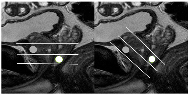
Figure 9.
Schematic figures illustrating the different scan axes between MRI and TRUS: (left figure) and (right figure) figures indicate the scan axes (white lines) of MRI and TRUS, respectively. The MRI and TRUS scan axes are placed in the perpendicular and oblique direction to the prostate urethra, respectively. On MRI, an anterior lesion (gray circle) is located between mid-gland and base, while on TRUS, it is seen between mid-gland and apex. On MRI, a posterior lesion (white circle) is located between mid-gland and apex, while on TRUS, it (white circle) is seen between mid-gland and base.

