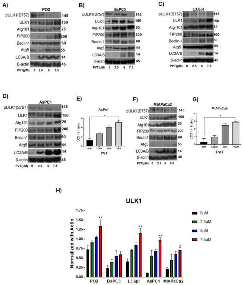
Figure 4.
PVT elevates ULK1 mediated autophagy in PDAC cells. Western blot analysis of pULK1(S757), ULK1, Atg101, FIP200, Beclin-1, Atg5, and LC3A/B in (A) PO2 (B) BxPC3 (C) L3.6pl, (D) AsPC1, and (F) MIAPaCa2 cells treated with 2.5, 5 and 7.5 µM PVT for 24 h. β-actin was used as a loading control. Figures shown are representative blots of three independent experiments. Blots were developed using Optimax X-ray film processor (Protec, Germany). PVT enhanced LC3 II/I ratio in (E) AsPC1 and (G) MIAPaCa2 cells treated with PVT for 24 h. Proteins were probed for LC3 antibodies and LC3 II/I band density was measured using Image J. (H) Quantitated representation of ULK1 expression data obtained from Figure 4A–D,F. * p ≤ 0.05, ** p ≤ 0.01.

