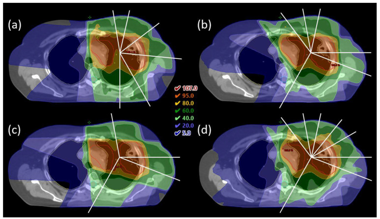
Figure A1.
(a) CLIN plan with manually selected beam angles for patient 4, with a frontal tumor and lymph nodes. (b–d) iCE plans with 6, 4 and 8 optimized beam angles for the same patient. Dose is shown relative to the prescribed dose (66 Gy). Contours are the PTV (red), lungs (yellow), esophagus (grey) and spinal cord (cyan), and beam angles are indicated by white lines. For this patient, the optimized beam angles in the 6- and 8-beam plans enabled sparing of the esophagus.

