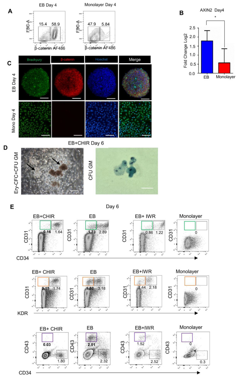
Figure 4.
Endogenous activity of Wnt/β-catenin signaling in 3D/EB and 2D/Monolayer cells on day 4 of PSC differentiation. (A) Representative flow cytometry analysis of Day 4 cells, comparison of β-catenin-positive cells determined by FACS, and (B) Quantitative PCR analysis of Axin2, and (C) Immunofluorescence of Brachyury and β-catenin. Scale Bars: 100 μm, (Mean ± SEM) n = 3, * p ≤ 0.05. (D) Phase-contrast images of disaggregated 3D/EB+CHIR day 6 cells cultured in methylcellulose; arrows indicate colonies of EryCFC and CFU-GM wright-stained. (E) Representative flow cytometry analysis of Day 6 3D/EB cells after the use of Wnt activator/inhibitor showing the results of the treatment either with CHIR or with IWR, green rectangle shows the population of interest, the endothelial component in CD31/CD34 cells, orange rectangle shows the endothelial component in the CD31/KDR cells and purple rectangle distinguishes the primitive hematopoietic component in the CD43/CD34 cells. Source of the cells: CD34/KDR populations (D), CD31/CD34, KDR/CD31, and CD34/CD43 populations.

