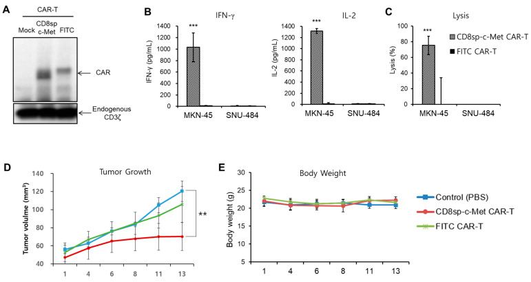
Figure 5.
c-Met CAR T cells effectively kill MKN-45 both in vitro and in vivo. T cells were activated with CD3/CD28 Dynabeads and were cultured in medium containing rhIL-7 and rhIL-15. Two days post-activation, T cells were transduced with c-Met CAR or FITC CAR lentivirus. (A) At 9 days post-activation, cell lysates were prepared for Western blot to see CAR expression. (B) CD8sp-c-Met CAR or FITC CAR T (2 × 10⁵ cells) were incubated with MKN-45 or SNU-484 (2 × 10⁴ cells). The next day, supernatants were collected to perform IFN-γ and IL-2 ELISA. (C) CD8sp-c-Met CAR or FITC CAR T (1.2 × 10⁵ cells) were co-incubated with MKN-45 or SNU-484 (1.5 × 10⁴ cells) at E:T ratio 8:1 for 20 h. The bars show mean ± S.D. of experiments performed in triplicate. Statistical analysis was performed by the paired t-test. ** p ≤ 0.05; *** p ≤ 0.01, compared with the FITC CAR-T group. (D,E) After irradiating 3 Gy to NSG (NOD.Cg-PrkdcscidIl2rgtm1Wjl/SzJ) mice, MKN-45 cells were implanted subcutaneously into the right flank of mice. Nine days after tumor implantation, control (PBS, n = 5), CD8sp-c-Met CAR T (n = 5), or FITC CAR T (n = 5) cells were intratumorally administrated. (D) Tumor volume and (E) weight of mice were assessed at 2–3-day intervals. The bars show mean ± S.E. of the experiment performed in 5 mice. Statistical analysis was performed by one-way ANOVA followed by Dunnett’s multiple comparisons test. ** p ≤ 0.05; *** p ≤ 0.01, compared with the control (PBS) group. (The whole western blots figure see Figure S1).

