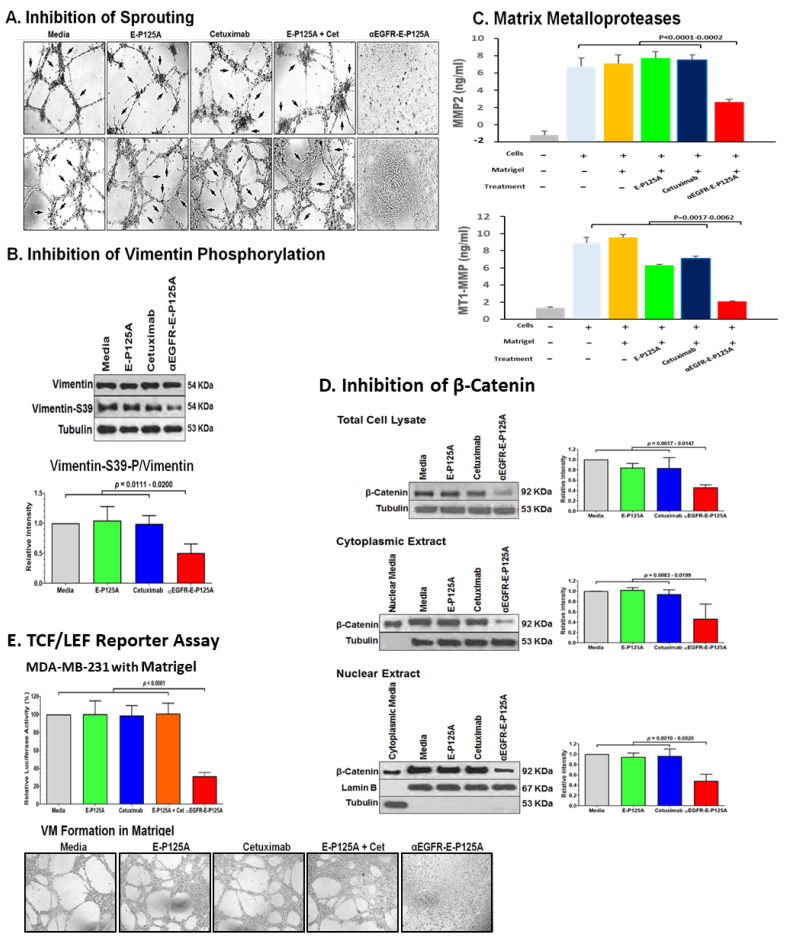
Figure 3.
(A) Effects of αEGFR-E-P125A on sprouting: Tube formation by HUVECs or MDA-MB-231-4175 plated on matrigel, untreated or treated with E-P125A (58 μg/mL), cetuximab (170 μg/mL, Cet), a combination of E-P125A (58 μg/mL) and cetuximab (170 μg/mL), or αEGFR-E-P125A (250 μg/mL) for 16 h. Sprouting indicated by black arrows. (B) Western blot analysis for vimentin and SER39-phosphorylated vimentin following αEGFR-E-P125A treatment: MDA-MB-231-4175 cells plated in matrigel were treated with E-P125A (58 μg/mL), cetuximab (170 μg/mL), or αEGFR-E-P125A (250 μg/mL) for 16 h and harvested; cytoplasmic extracts were prepared, and western blot performed for vimentin or SER39-phosphorylated vimentin. Relative quantification of vimentin-S39-P/vimentin using the NIH ImageJ program is shown. (C) αEGFR-E-P125A inhibits secretion of MMP2 and shedding of MT1-MMP: MDA-MB-231-4175 cells were plated on matrigel, treated with E-P125A (58 μg/mL), cetuximab (170 μg/mL), or αEGFR-E-P125A (250 μg/mL) for 16 h; supernatants were harvested and analyzed for secreted MMP2 or shed MT1-MMP by ELISA. Details in methods. (D) αEGFR-E-P125A treatment reduces β-catenin levels. MDA-MB-231-4175 cells were plated on matrigel, treated with E-P125A (58 μg/mL), cetuximab (170 μg, or αEGFR-E-P125A (250 μg/mL) for 16 h and then harvested; total, cytoplasmic, and nuclear extracts were analyzed by Western blotting. Tubulin was used for cytoplasmic and lamin B for nuclear normalization. The right histogram shows the relative quantification of β-catenin/(tubulin or lamin B) staining using NIH ImageJ software. (E) αEGFR-E-P125A inhibits TCF/LEF transcriptional activity. MDA-MB-231 (TCF/LEF Luc Reporter) cells were plated and treated as indicated (Figure 3A). TCF/LEF reporter assay was performed in 96-well plates without (2D) or containing matrigel (3D, shown in the bottom panel) at 16 h. Luciferase activity in treated cells relative to the medium control is indicated.

