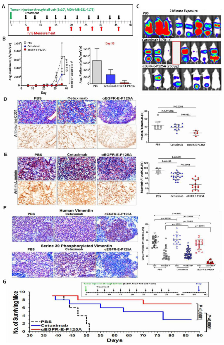
Figure 5.
(A–F) αEGFR IgG1-huEndo-P125A inhibits pulmonary tropic MDA-MB-231-4175 lung metastasis. (A) Pulmonary metastasis: 5 × 10⁴ MDA-MB-231-4175 cells were injected via the tail vein (green arrow). Mice (n = 8) were treated starting day 5 with PBS, cetuximab (170 µg/injection), or αEGFR-E-P125A (250 µg/injection) 2×/week (black arrows, 9 injections). Metastases were monitored by IVIS at intervals indicated by red arrows. Experiment terminated on day 36. A single mouse was lost in the cetuximab group on day 35 (a red blank in Figure 5C). (B,C) Pulmonary average photon flux graph (B) and relative photon flux/2-min exposure on day 36 (C) The αEGFR-E-P125A-treated group showed reduced total body tumor burden and lung metastasis. Average photon flux on day 36 is shown in a graph. Data shown as the mean ± SEM. (D,E) αEGFR-E-P125A inhibits mCD31+ angiogenesis (D) and huLaminin+ TNBC VM (E) of MDA-MB-231-4175. Immunohistochemical staining of tumor sections on day 36 from mice in Figure 5B and Figure 6C. Cryosections from representative treated mice were stained with anti-murine CD31 (D) or anti-human laminin antibody (E) and counterstained with hematoxylin. Relative positive optical density/total optical density of the immunohistochemical staining was analyzed using NIH ImageJ. Each dot represents an immunohistochemically stained cryosection from a tumor selected for near ‘average’ photon flux intensity. Details in Methods. (F) αEGFR-E-P125A inhibits vimentin-SER39 phosphorylation: On day 36, lung sections containing metastatic MDA-MB-231-4175 from sacrificed mice (Figure 5B,C) were analyzed by IHC. Cryosections from mice with near average relative photon flux intensity stained for human vimentin (brown, Vim, the upper panel) or human vimentin phosphorylated on serine 39 (brown, VimS39-P, bottom panel). Hematoxylin was used for nuclear counter-staining. Representative tumor cryosections from mice treated with PBS, cetuximab, or αEGFR-E-P125A are presented. Relative vimentin+ or SER39 vimentin+ staining optical density/total optical density of IHC staining was quantified using NIH ImageJ. (G) Survival of mice following αEGFR-E-P125A treatment. MDA-MB-231-4175 cells (5 × 10⁴) were injected via the tail vein and survival was measured. Nine mice/group were treated starting day 5 with PBS, cetuximab (68 µg/injection), or αEGFR-E-P125A (100 µg/injection) 2×/week (10 injections) per schedule shown above the survival curve. Survival was analyzed using the survival analyses and one-way ANOVA test (GraphPad Prism 7.03). Green arrow, tumor inoculation; black arrow: treatment administration ( PBS, cetuximab, or αEGFR-E-P125A as indicated), blue arrow: experiment termination.

