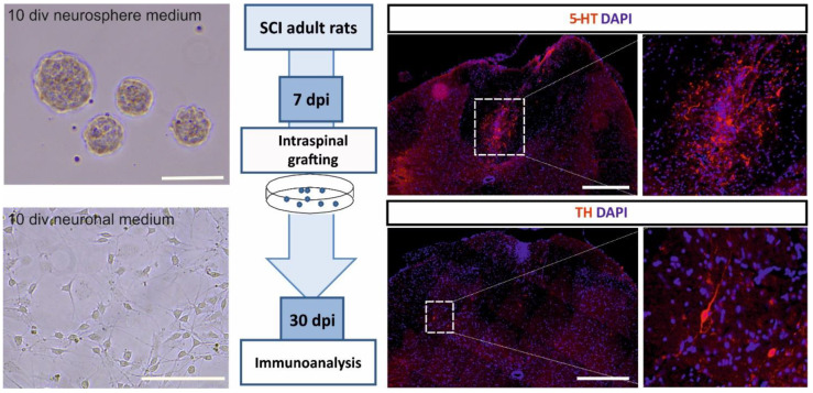
Figure 2.
Schematic presentation of the results of preliminary experiments showing serotonergic precursors surviving and terminally differentiating after transplantation into the spinal cord injury-mediated environment. The left panel shows precursor morphology in different cell culture conditions: Neurosphere medium supports cell proliferation and neurosphere formation, neuronal medium induces their neuronal differentiation. Scale bars 100 μm. The middle panel describes the experimental design: Expanded serotonergic precursors derived from rat embryonic brain stem (on embryonic day 11) were transplanted into the rat injured spinal cord 7 days post-injury and their morphology was examined after the next 30 days. The protocol for cell expansion was adopted from [196]. The right panel shows representative images of differentiated cells within host spinal cords. Transplant-derived serotonergic neurons were found in each examined injured spinal cord (three rats). Note the presence of serotonin (5-HT)-positive neurons (upper images) and thyrosine kinase (TH)-positive neurons (lower images). Control animals were injected with cell culture medium, no stained cells were detected (data not shown). Scale bars are 500 μm. Functional experiments that examined the recovery of locomotor functions via restored innervation of the spinal cord are in progress.

