Figure 1.
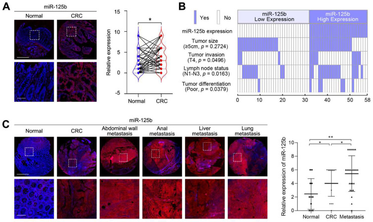
miR-125b expression is upregulated in metastatic colorectal cancer (CRC) tissues. (A) Representative images and analysis of fluorescence in situ hybridization (FISH) staining for miR-125b in 58 cases of CRC tissues and their matched adjacent normal tissues. p = 0.0130 (n = 58; Normal vs. CRC). Magnified views of the regions indicated by the boxed area are shown below. Scale bars represent 200 μm (upper) and 100 μm (lower). (B) miR-125b expression positively correlated with tumor invasion, lymph node metastasis and tumor differentiation. (C) Representative images and analysis of miR-125b expression in normal colon tissues, primary CRC tissues and metastatic tissues detected by FISH. p = 0.0489 (Normal vs. CRC); p = 0.0423 (CRC vs. Metastasis); p = 0.0025 (Normal vs. Metastasis); n (Normal) = 11; n (CRC) = 21; n (Metastasis) = 25. Scale bars represent 500 μm (upper) and 100 μm (lower). * p < 0.05, ** p < 0.01 by Student’s t-test.

