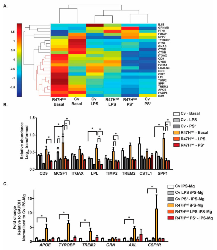
Figure 2.
Proteins associated with the DAM microglia signature are found in exosomes. The exosomal proteomic profile was compared with a list of published DAM-related proteins [36,37,38]. R47Hhet exosomes contained a subset of DAM-associated proteins (indicated in orange) at higher levels than Cv exosomes (A). These proteins were particularly enriched with the TREM2-dependent second stage of DAM [36]. The relative abundance of the DAM proteins in the exosomes was normalised to the abundance observed in Cv exosomes of these second-stage DAM-associated proteins (B). The expression of DAM-associated genes was measured in the iPS-Mg using qPCR to assess whether the changes observed in the exosomes reflected differences at the cellular level or in the packaging (C). For (A,B), N = 3 independent samples analysed through LC-MS/MS for each condition, whilst for (C), N = 4 independent experiments. Two-way ANOVA with * p < 0.05, ** p < 0.01.

