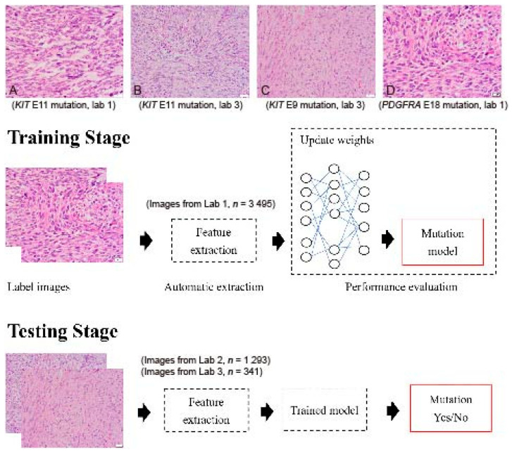
Figure 1.
Flowchart of the implemented deep convolutional neural network. Images were collected from 3 independent laboratories (Lab 1, Lab 2, and Lab 3) using 4 µm thick, hematoxylin-and-eosin slides. (A) An example image of a GIST with a KIT exon 11 mutation from Lab 1 (400×). (B) An example image of a GIST with a KIT exon 11 mutation from Lab 3 (400×). (C) An example image of a GIST with a KIT exon 9 mutation from Lab 3 (400×). (D) An example image of a GIST with a PDGFRA exon 18 mutation from Lab 1 (400×). Although similar staining protocols were used, the overall hue differs between laboratories. A total of 5129 images were enrolled in the study. Four different DCNNs, AlexNet, InceptionV3, ResNet, and DenseNet, were used with transfer learning from ImageNet. The transferred DCNN model generated the probability that each image contained a drug-sensitive mutation. A probability higher than 0.5 was regarded as positive. The dataset was randomly separated into 10 equal-sized subsets. In each iteration, one subset was picked and used to test the trained model on the remaining nine subsets.

