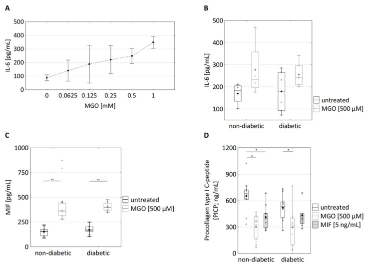
Figure 3.
Effects of MGO and MIF treatment of dermal fibroblasts on the secretion of immunomodulatory cytokines and procollagen type I C-peptide. (A) Cultured dermal fibroblasts were treated with the indicated MGO concentrations (ranging from 0.0625 to 1 mM) for 24 h. The supernatants were analysed for IL6 levels using Homogeneous Time Resolved Fluorescence assay n = 6 (3 non-diabetic, 3 diabetic). (B) Cultured fibroblasts from diabetic and non-diabetic donors were treated with 500 µM MGO for 24 h. The supernatants were analysed for IL6 levels using Homogeneous Time Resolved Fluorescence assay; nnon-diabetic = 5; ndiabetic = 7. (C) The supernatants of cultured fibroblasts from diabetic and non-diabetic donors were analysed for MIF using ELISA. They were treated with 500 µM MGO for 24 h; nnon-diabetic = 9; ndiabetic = 10. (D) The supernatants were analysed for procollagen type I C-peptide using ELISA. Fibroblasts from diabetic and non-diabetic donors were treated with 500 µM MGO and 10 ng/mL MIF for 24 h; nnon-diabetic = 9; ndiabetic = 10. Statistical significance was determined by Mann–Whitney-U-Test between groups and by Wilcoxon test for comparison between untreated and treated; * p < 0.05, ** p < 0.01.

