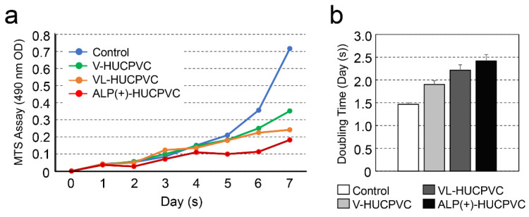
Figure 4.
Change in cell proliferation of HUCPVCs. (a) Changes in cell proliferation over time by MTS assay. V-HUCPVCs, VL-HUCPVCs, and ALP(+)-HUCPVCs were cultured at a final volume of 120 μL/well for 1 h at 37 °C. MTS reagent was added, and absorbance of 490 nm was recorded using a microplate reader. (b) Cell population doubling level against days. Control: HUCPVCs cultured in the absence of VD, LDN, and TGF-β1.

