Figure 5.
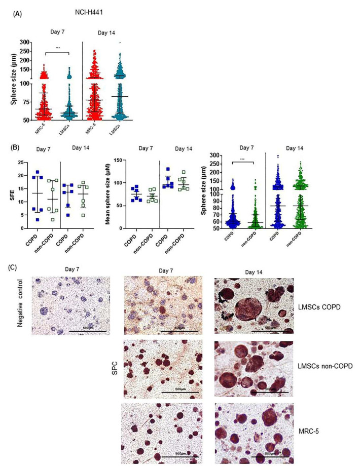
Abnormalities in COPD-derived lung-resident MSCs (LMSCs)-supported organoid formation by NCI-H441 cells. NCI-H441 cells together with mitomycin-treated MRC-5 cells or mitomycin-treated LMSCs from COPD and control donors were seeded into 100 μL growth factor-reduced 1:1 diluted Matrigel onto inserts of a transwell and cultured for 7–14 days. The number per well and the size of organoids were measured using light microscopy. The sphere forming efficiency (SFE) was determined as number of organoids normalized by cell input. (A) Size of organoids generated in the presence of LMSCs (n = 12, COPD and control donors combined) or MRC-5 cells. Medians ± IQR are indicated. (B) SFE, mean size and size distribution of organoids generated in the presence of COPD (n = 6) and non-COPD (n = 6) LMSCs. Medians ± IQR are indicated. (C) Inserts were stained with for SPC (using AEC and HE counterstaining). Representative images are shown of 2 independent experiments with 3 non-COPD and 3 COPD LMSC donors per experiment. *** = p < 0.001, * = p < 0.05 between the indicated values as analyzed by the Mann Whitney U test.

