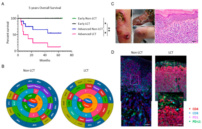
Figure 1.
Kaplan–Meier analysis shows significantly decreased survival of patients with LCT-MF compared to non-LCT-MF. (A) The overall survival of early non-LCT (green) or early LCT-MF (purple) patients was compared to advanced non-LCT (blue) or advanced LCT-MF patients (magenta) and determined by Mantel–Cox test with p value < 0.05 considered significant (n = 28). * p < 0.05, ** p < 0.01. (B) Circular charts show the distribution of demographic characteristics including stage, MF subtype, gender, race, and survival status of LCT and non-LCT patients. (C) Clinical presentation of patients with LCT-MF and Hematoxylin and Eosin (H&E) staining of the LCT-MF lesional skin. Scale bar = 50 μm. (D) Representative multiplex immunofluorescence images of CTCL skin biopsy with LCT-MF and non-LCT-MF. CD4 is stained red; CD8 is blue; PD1 is magenta; PD-L1 is green. Scale bar = 20 μm.

