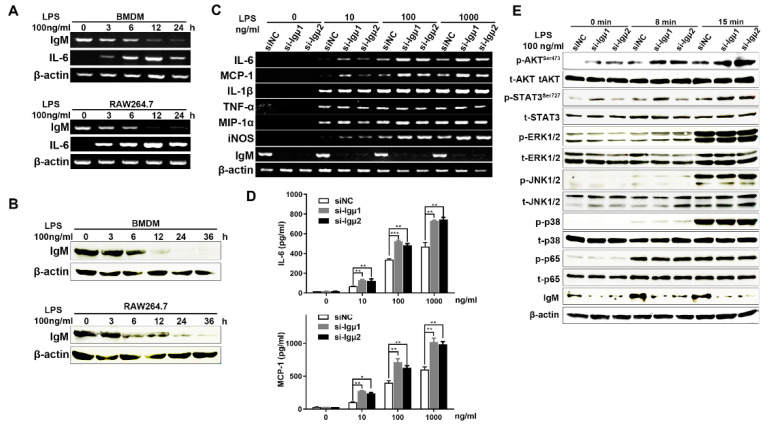
Figure 4.
IgM knockdown promoted a pro-inflammatory effect in LPS-induced RAW264.7 macrophages through Akt signaling. The upregulated expression of IgM in BMDMs and RAW264.7 cells stimulated with 100 ng/mL LPS for 0, 3, 6, 12, and 24 h by RT-PCR (A) and Western blot (B). (C) Cells were transfected with IgM siRNA (si-Igμ) and control siRNA (siNC) for 36 h and treated with various concentrations of LPS (0, 1, 10, 100, and 1000 ng/mL) for an additional 3 h. The pro-inflammatory cytokines, including IL-6, MCP-1, IL-1β, TNF-α, MIP-1α, and iNOS, were determined by RT-PCR. IgM acted as positive control and β-actin as an internal control. (D) Cells were transfected with IgM siRNA (si-Igμ) and control siRNA (siNC) for 36 h and treated with LPS (0, 1, 10, 100, and 1000 ng/mL) for an additional 12 h. The production of IL-6 and MCP-1 cytokines was measured by ELISA kit using the microplate reader. The data are presented as means ± SD (n = 3). (* p < 0.05 and ** p < 0.01, *** p < 0.001 vs. siNC control group). (E) Cells were transfected with IgM siRNA (si-Igμ) and control siRNA (siNC) for 36 h before exposure to LPS (100 g/mL) for 8 and 15 min. Enhanced effects of IgM knockdown on phosphorylation of AKT at Ser 473 and STAT3 at Ser 727 in LPS-induced RAW264.7 macrophages were shown by Western blot analysis, but no obvious effect on phosphorylation of ERK1/2, JNK1/2, p38, and p65 was shown. p, phosphorylated protein; t, total protein. IgM acted as positive control and β-actin as internal control.

