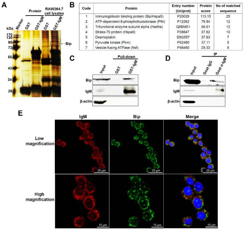
Figure 5.
IgM interacted with Bip in RAW264.7 cells. (A) Whole cell lysate derived from RAW264.7 cells was used to pull down with the same amount of GST-IgM recombinant protein or GST protein as control, respectively. The complexes were separated on SDS-PAGE gels. After silver staining, the differential bands or dots were analyzed by MS. Asterisks and circles indicated for the differential band of Bip. (B) Candidates of IgM-associated proteins were identified by liquid chromatography-mass spectrometry. Only part of the identified proteins was shown after removal of repeated proteins and proteins related to the heavy or light chains of the antibody. (C) Western blot analysis to determine the interaction between IgM and Bip in GST-pull down assay. (D) The interaction between endogenous IgM and Bip was shown in the coimmunoprecipitation assay. The cell lysate of RAW264.7 cells was immunoprecipitated with goat anti-IgM antibody and goat IgG as control. Input, RAW264.7 cell lysate. (E) Confocal microscopy analysis of IgM (red) and Bip (green) in RAW264.7 cells using goat anti-mouse IgM polyclonal antibody and rabbit anti-mouse Bip antibody. DAPI (blue) was used for nuclear staining. Scale bars, 25 μm (Low magnification) and 10 μm (High magnification).

