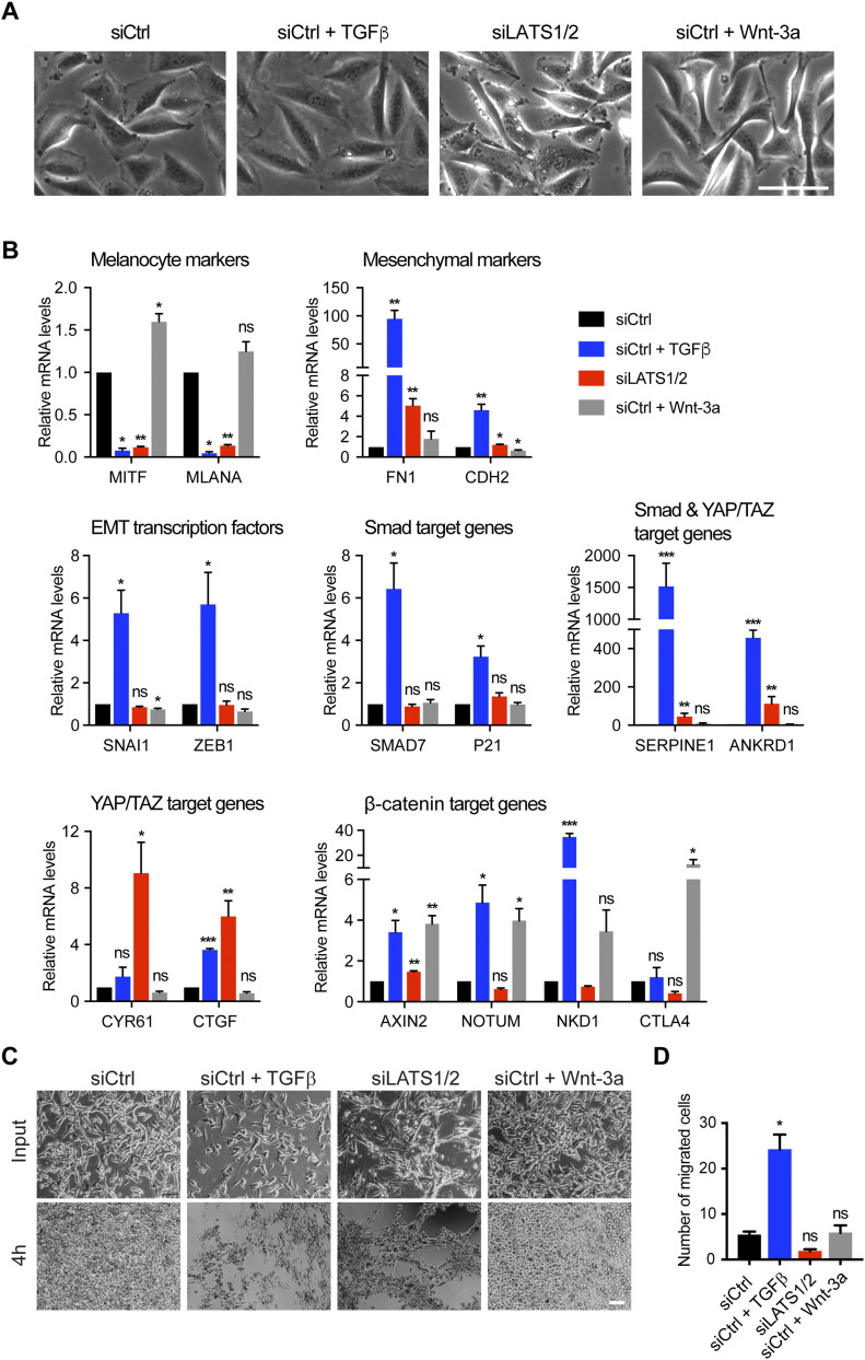
Figure 1. TGFβ/SMAD and YAP/TAZ induce an invasive cell phenotype, whereas Wnt/β-catenin promotes a proliferative cell phenotype in proliferative-type melanoma cells.
(A) Representative phase-contrast images of proliferative-type M000921 cells treated with TGFβ, siLATS1/2, or Wnt-3a for 2 d. Scale bar, 100 μm. (B) Quantitative RT-PCR analysis of the expression of melanocyte marker genes (MITF and MLANA), mesenchymal marker genes (FN1 and CDH2), EMT transcription factors (SNAI1 and ZEB1) and SMAD (SMAD7 and P21), SMAD/YAP/TAZ (SERPINE1 and ANKRD1), YAP/TAZ (CYR61 and CTGF), and β-catenin target genes (AXIN2, NOTUM, and NKD1) in proliferative-type M000921 cells treated with siControl (siCtrl), siCtrl + TGFβ, siLATS1/2, or siCtrl + Wnt-3a for 2 d. Mean + SEM of n = 3 replicates are shown *P < 0.05; **P < 0.01; ***P < 0.001; ratio-paired t test. (C) Invasive growth in 3D Matrigel culture. Proliferative-type M010817 cells were transfected with siCtrl or LATS1/2 or treated with TGFβ or Wnt-3a for 5 d as indicated and subsequently cultured in diluted 3D Matrigel. Representative pictures were taken by phase-contrast microscopy after seeding and 4 h thereafter, revealing the morphological changes and invasive growth of TGFβ and siLATS1/2-treated cells in Matrigel. Scale bar, 100 μm. (D) Cell migration assay. Proliferative-type M010817 melanoma cells were transfected with siCtrl or siLATS1/2 or treated with TGFβ or Wnt-3a for 5 d and subsequently seeded in modified Boyden Chamber culture insets with 10% FBS as chemoattractant in the bottom well. Cells that have migrated after treatment with siCtrl, siLATS1/2, TGFβ, or Wnt-3a were quantified after 20 h. *P < 0.05; ratio-paired t test.

