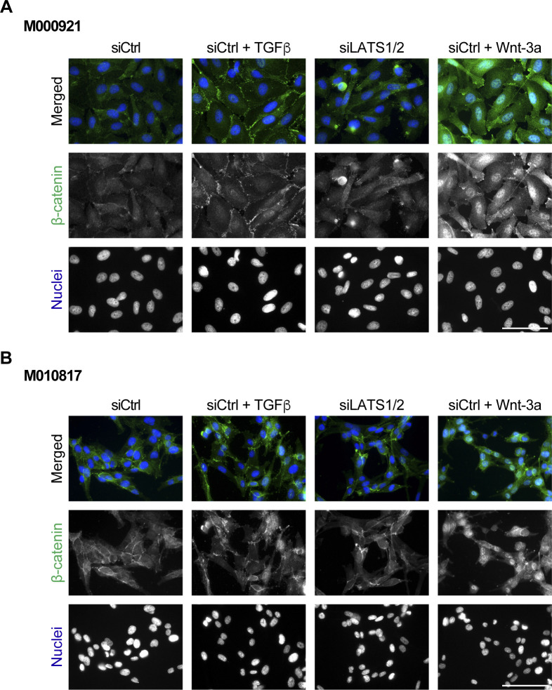
Figure S3. Nuclear localization of β-catenin upon stimulation with TGFβ, Wnt-3a, or siRNA-mediated ablation of LATS1/2 and subsequent YAP/TAZ activation.
(A, B) M000921 (A) and M010817 (B) proliferative melanoma cells were treated with siCtrl, siCtrl plus TGFβ, siLATS1/2, and siCtrl plus Wnt-3a, and the nuclear localization of β-catenin was assessed by immunofluorescence staining. DAPI was used to visualize nuclei. Representative microscopy images of n = 2 are shown. Scale bars, 100 μm.

