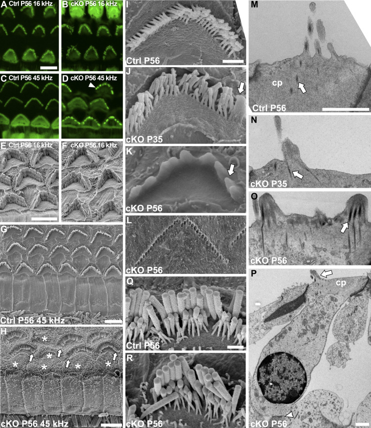
Figure 4. Adult Manffl/fl;Pax2-Cre B6 mice show progressive deterioration of the outer hair cell (OHC) hair bundles.
(A, B) At P56, comparison of phalloidin labeling in the 16-kHz region of mutant and control cochleas shows disorganized OHC hair bundles only in mutants (C, D) Phalloidin labeling in the 45-kHz region of mutant specimens shows severe bundle disorganization, in the form of bundle fragmentation (arrowhead). (E, F) Scanning electron microscopy (SEM) images from the 16-kHz region confirms the loss of cohesion of OHC bundles in mutant mice. (G, H) SEM images from the 45-kHz cochlear region show OHCs deterioration in cKO mice, such that most OHCs have fused stereocilia (arrows) and many of them are lost (*, site of lost OHC). (I, J, K) SEM views showing age-related progression in hair bundle deterioration in cKO mice. The bundles exhibit stereocilia fusion at their edges (arrow) at first, shown at P35. The fusion progresses and affects the entire bundle, shown at P56. (L) Stereocilia imprints of a high-frequency OHC embedded in the tectorial membrane in P56 mutant specimen are shown (see text for details). (M, N, O) Transverse transmission electron microscopy (TEM) sections of high-frequency OHCs from mutant and control mice. The sections from P35 and P56 mutant specimens confirm stereocilia fusion and show apical membrane lifting that creates this fusion. The rootlets (arrows) remain separate in the fused structures. (P) A transverse TEM section of a 45-kHz OHC from a P56 mutant specimen shows no signs of cell pathology other than the hair bundle fusion. Both efferent (arrowhead) and afferent (not shown) synaptic connections can be found. (Q, R) SEM images reveal a comparable appearance of 45-kHz IHC hair bundles in P56 mutant and control mice. Abbreviations: B6, C57BL/6J strain; cKO, conditional knock out; cp, cuticular plate; Ctrl, control; IHC, inner hair cell; OHC, outer hair cell; SEM, scanning electron microscopy; TEM, transmission electron microscopy. Scale bars: (A) 5 μm A-D; (E) 5 μm E, F; (G) 5 μm; (H) 5 μm; (I) 1 μm I-L; (M) 1 μm M-O; (P) 1 μm; (Q) 1 μm Q, R.

