. 2021 Nov 25;10:e69418. doi: 10.7554/eLife.69418

Figure 3. Structure of the M. tuberculosis cytochrome bcc subunits.

Cartoon representation of the monomers of (A) QcrA, (B) QcrB, and (C) QcrC, with prosthetic groups. (D) The QP-binding site and (E) QN-binding site. The residues potentially involved in the binding of MK/MKH2 are shown with side chains in stick model representation. MK/MKH2 have their carbon atoms in green and are represented as stick models. The [2Fe-2S] and heme groups are shown as spheres and labeled.

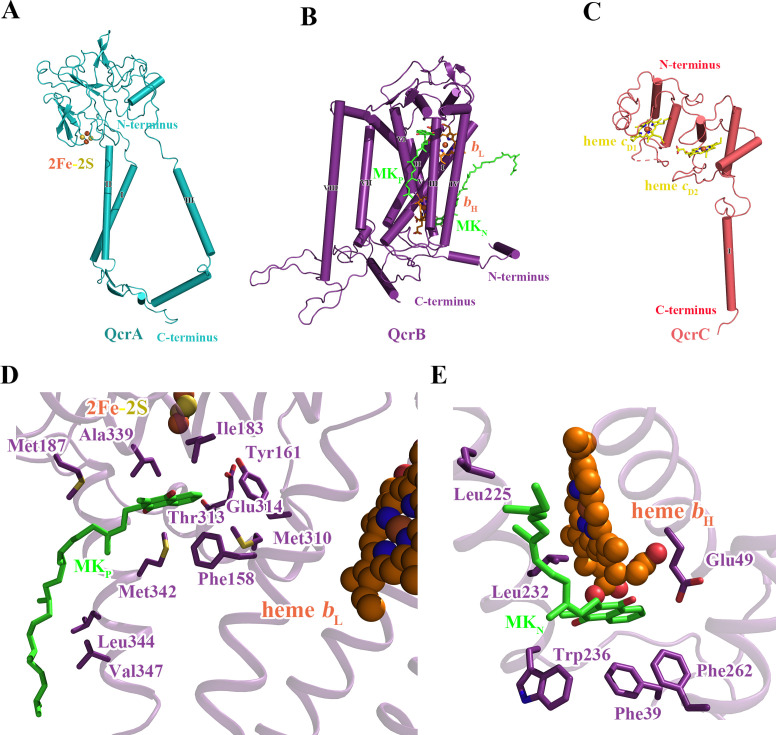
Figure 3.

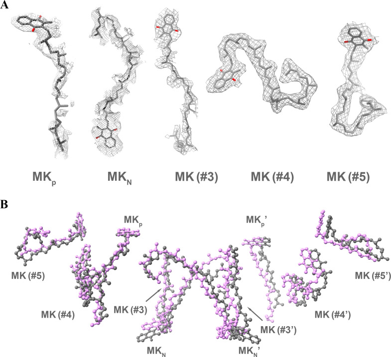
Figure 3—figure supplement 1. Identification and comparison of MK/MKH2 in the hybrid supercomplex.

Figure 3—figure supplement 1.

Ten MK/MKH2 (gray) have been assigned according to the densities (A) and are in the same locations as observed in the M. smegmatis CIII2CIV2 supercomplex (magenta) (PDB 6ADQ) (B).
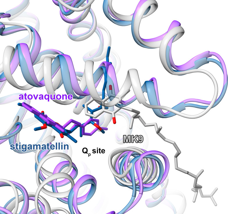
Figure 3—figure supplement 2. Comparison of M. tuberculosis CIII quinol-binding site (gray) with those from CIII from S. cerevisiae (PDB 3CX5) (blue) and (PDB 4PD4) (purple).

Figure 3—figure supplement 2.

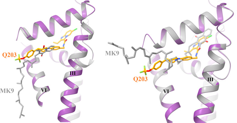
Figure 3—figure supplement 3. Structural superposition of native (left) and docked (right) MK9 with the Q203 in the Qp site.

Figure 3—figure supplement 3.

The QcrB is extracted from the hybrid supercomplex with Q203 (purple) and MK9 (gray) bound is shown in cartoon representation.