. 2021 Nov 25;10:e69418. doi: 10.7554/eLife.69418

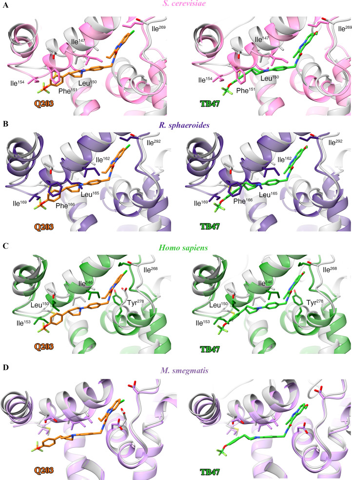
Figure 6. Structural alignment between the M. tuberculosis Qp-binding pocket where Q203 or TB47 binds with homologous subunits from four other species.

These subunits are from (A) S. cerevisiae (pink, PDB: 1KYO), (B) R. sphaeroides (blue, PDB: 2QJP), (C) Homo sapiens (green; PDB: 5XTE), and (D) M. smegmatis (violet, PDB: 6ADQ). Residues causing steric clashes in the homologous subunits are labeled.

Figure 6.

Figure 6—figure supplement 1. Sequence alignment of M. tuberculosis QcrB with their counterparts in other species including Homo sapiens.
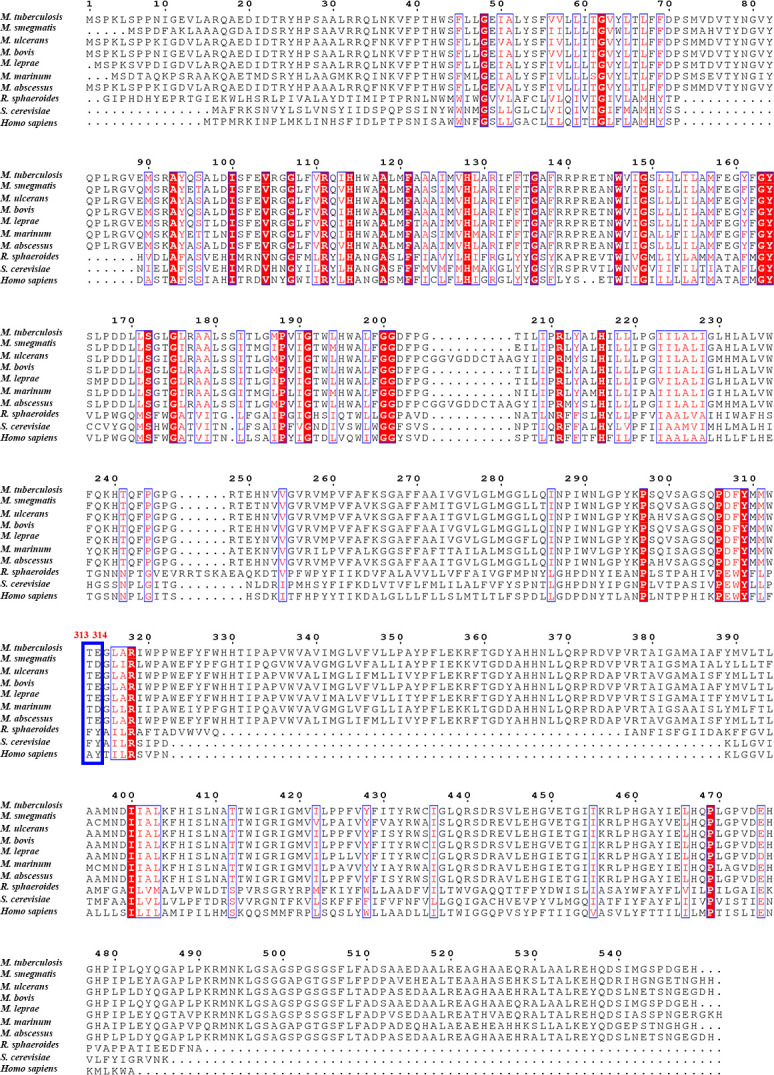
Figure 6—figure supplement 1.

Red residues are conserved, and blue indicates those less well conserved.
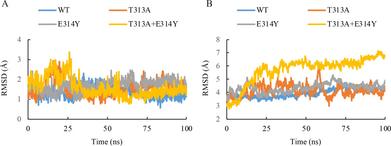
Figure 6—figure supplement 2. Molecular dynamics simulation plot for the root mean squared deviation (RMSD) of the heavy atoms in the inhibitor (A) and main chain atoms of QcrB (B).

Figure 6—figure supplement 2.

100 ns NPT simulations for the wild-type and three mutant systems of the Q203-bound QcrB complex (T313A, E314Y, T313A + E314Y) are recorded. The frames were extracted from the 100 ns simulation every 100 ps, generating 1000 frames. The RMSD values of each frame were calculated based on the structural conformation of the reference structures, namely the initial wild-type and mutant Q203-bound QcrB complexes, respectively.
Figure 6—figure supplement 2—source data 1. Root mean squared deviation of the inhibitor and protein of Q203-bound QcrB complex.