. 2021 Nov 25;10:e69418. doi: 10.7554/eLife.69418

Figure 7. Schematic of M. tuberculosis cytochrome bcc inhibition by Q203 and TB47.

The two monomers of M. tuberculosis cytochrome bcc are colored magenta and cyan, respectively. The binding of Q203 (orange spheres) or TB47 (green spheres) prevents substrate access (gray spheres).

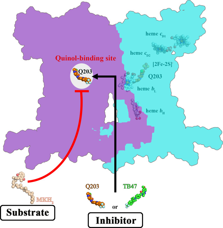
Figure 7.

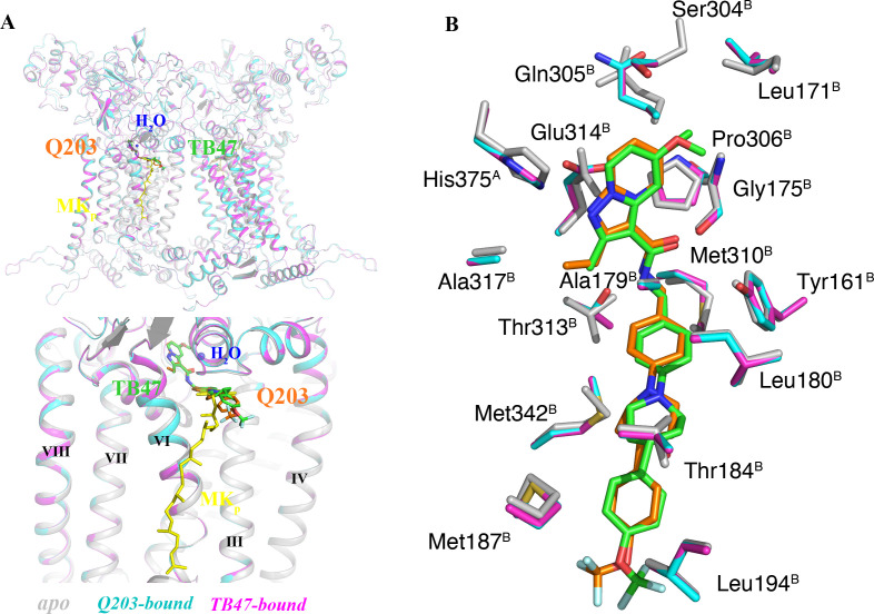
Figure 7—figure supplement 1. Comparison of apo and Q203/TB47-bound structures of M. tuberculosis cytochromes bcc.

Figure 7—figure supplement 1.

(A) Superposition of apo (gray), Q203-bound (cyan), and TB47-bound (magenta) structures of M. tuberculosis cytochromes bcc. The Q203 (orange), TB47 (green), and MK (yellow) molecules are shown as stick models, respectively. Water molecules are shown as spheres. (B) Comparison of residues surrounding Q203 (cyan sticks) and TB47 (magenta stick models) with those in apo form (gray sticks). The residues from subunits A and B are labeled with superscript A and B, respectively.
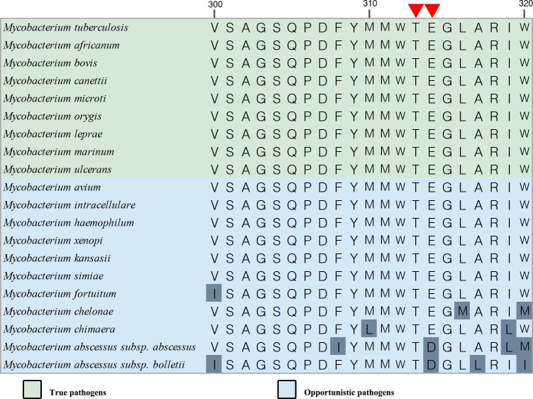
Figure 7—figure supplement 2. Sequence alignment for the QP site within pathogenic mycobacteria.

Figure 7—figure supplement 2.

Partial sequence alignment at the Qp site. The red triangles indicate the conservative residues involved with the hydrogen-bond formation between Q203 and TB47.