Figure 5.
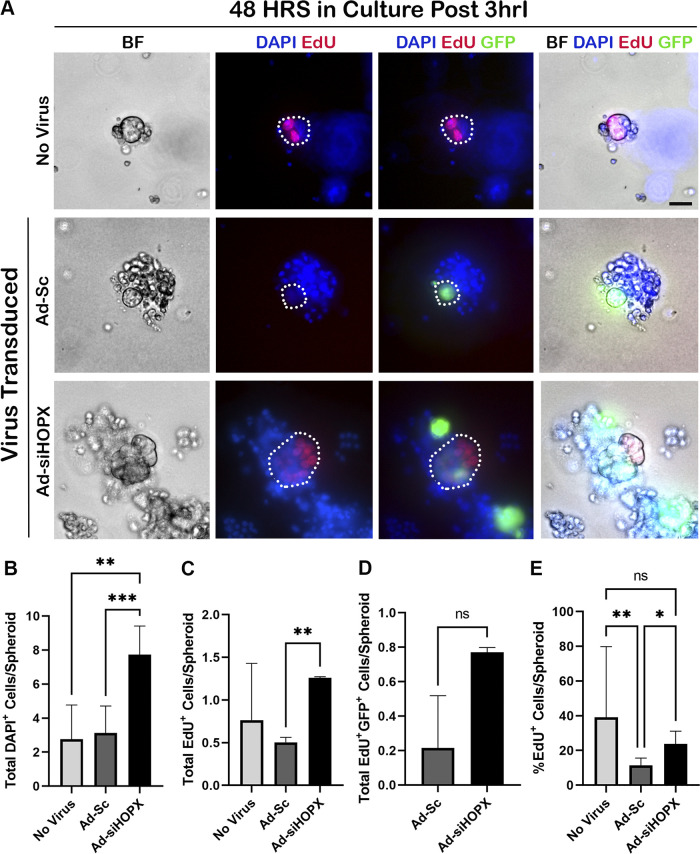
Silencing HOPX in 3-h ischemic injured (3hrI) crypt epithelium leads to increased overall cell and proliferating cell counts at 48 h in culture. A: representative bright field (BF) and immunofluorescent images of 48 h spheroids from no virus, Ad-Sc, and Ad-siHOPX-transduced 3hrI crypts. Proliferating cells were identified by EdU (red) and Ad-Sc and Ad-siHOPX cells identified by their GFP reporter (green). Nuclei were stained with Hoescht 33342 (blue). Scale bar 20 µm. B: means and standard deviations of total cell count per spheroid, showing significantly increased total cell counts for Ad-siHOPX compared with both Ad-Sc and no virus. C: means and standard deviations of total EdU+ cell counts per spheroid, showing significantly increased EdU+ cells in Ad-siHOPX compared with Ad-Sc. D: means and standard deviations of total EdU+GFP+ colocalized cells per spheroid, showing no difference between Ad-siHOPX and Ad-Sc. No virus spheroids are not included as they did not contain a GFP reporter. E: means and standard deviations of the percentage of EdU+ cells per total cells per spheroid, showing that with Ad-siHOPX leads to significantly increased percentage of proliferating cells. Analysis by two-way ANOVA with post hoc Tukey’s test, *P < 0.05 **P < 0.01 ***P < 0.001, n = 1 or 2 pigs, 15–28 enteroids counted per pig.

