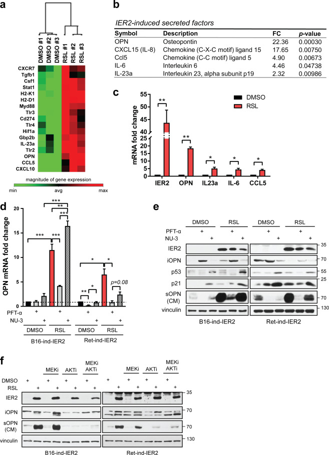
Fig. 4. Osteopontin is a major component of the p53-dependent IER2-induced SASP.
a Heatmap showing the top IER2-induced genes in B16-ind-IER2 #51 cells treated either with DMSO or RSL (3 × 0.5 μM) assessed using a Mouse Cancer Inflammation and Immunity Crosstalk PCR Array. b IER2-induced genes based on the qRT-PCR analysis (CXCL15, IL-6) and the Mouse Cancer Inflammation and Immunity Crosstalk PCR Array (OPN, Ccl5, and IL23a). Only genes with a fold change (FC) ≥ 2.0 and p < 0.05 are shown. See also Table S3. c qRT-PCR quantification of IER2, OPN, IL-23a, IL-6, and CCL5 in B16-ind-IER2 #51 cells treated with either DMSO or RSL (3 × 0.5 μM). RPLP0 was used for normalization. Bars represent means + SEM; n = 3. d qRT-PCR quantification of OPN in B16-ind-IER2 #51 and Ret-ind-IER2 #69 cells treated with PFT-α (20 μM) or NU-3 (5 μM) with or without stimulation with RSL (3 × 0.5 and 1 μM). RPLP0 was used for normalization in qRT-PCR. Bars represent mean + SEM; n = 3. e Western blot analysis of intracellular OPN (iOPN), IER2, p53, p21, vinculin (loading control), and secreted OPN (sOPN) present in conditioned media (CM) of B16-ind-IER2 #51 and Ret-ind-IER2 #69 cells treated with PFT-α (20 μM) or NU-3 (5 μM), with or without stimulation with RSL (3 × 0.5 and 1 μM). f Western blot analysis of intracellular OPN (iOPN), IER2, vinculin, and secreted OPN (sOPN) present in conditioned media (CM) of B16-ind-IER2 #51 and Ret-ind-IER2 #69 cells treated with or without RSL (3 × 0.5 and 0.5 μM) in the presence or absence of MEKi and/or AKTi (both 3 × 1 μM). Statistical significance was determined using an unpaired Student’s t test. *p < 0.05, **p < 0.01.

