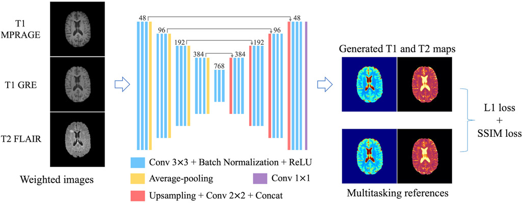
FIGURE 1.
Network design. A 2D U-Net-based architecture with four down-sampling steps and four up-sampling steps was used. 2×2 average pooling layers were used in the down-sampling track. Bilinear interpolation-based up-sampling layers followed by 2×2 convolution were used in the up-sampling track. At each resolution scale, 3 convolutional groups were sequentially applied, each consisting of 3×3 convolution followed by batch normalization and rectified linear unit (ReLU). Long skip connections were added to recover fine details in the up-sampling track. Slices of the three conventional weighted images (i.e., T1 MPRAGE, T1 GRE and T2 FLAIR) were concatenated as the 3-channel input. Slices of T1 and T2 maps formed the 2-channel target. A combination of L1 loss and SSIM loss was used as the loss function.

