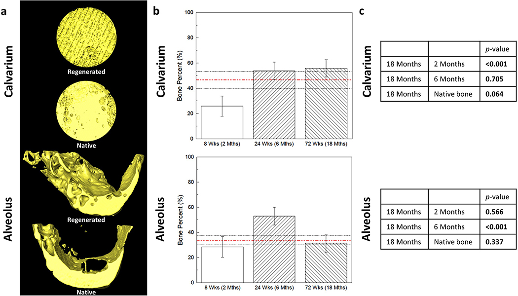
Figure 2.
(a) 3D reconstructions of scaffold-regenerated bone (yellow) at 18 months in vivo demonstrate regeneration across entire span of calvarial and alveolar defects, with morphology comparable to contralateral un-operated native bone. (b) Volumetric analysis of shows comparable bone volume fraction regenerated by scaffold compared to native bone (red dashed line; black dashed lines are 95% confidence intervals) at 18 months in both (c) calvarium and alveolus (p = 0.064 and p = 0.337, respectively). Error bars are 95% confidence intervals. Bone Percent = Bone Volume/Total Volume.

