
FIGURE 4.
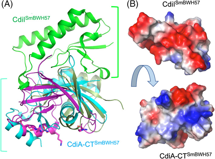
The crystal structure of CdiA‐CTSmBWH57•CdiISmBWH57 complex. (A) The toxin component (cyan) is compared with the RNase domain of histidine‐free ribonuclease colicin E5 (magenta) and BrnT toxin from Brucella abortus (dark green). The C‐terminal domain of CdiA‐CT covers approximately half of the RNase protein. The key residues in the active site of colicin E5 are shown as sticks. There are no equivalent residues in CdiA‐CTSmBWH57. The CdiISmBWH57 (green) does not have any close structural homologs in PDB. The CdiISmBWH57 interacts tightly with toxin using different surface on opposite site of colicin E5 active site. (B) Interacting surfaces of CdiA‐CTSmBWH57and CdiISmBWH57 are highly complementary in terms of shape and charge potential