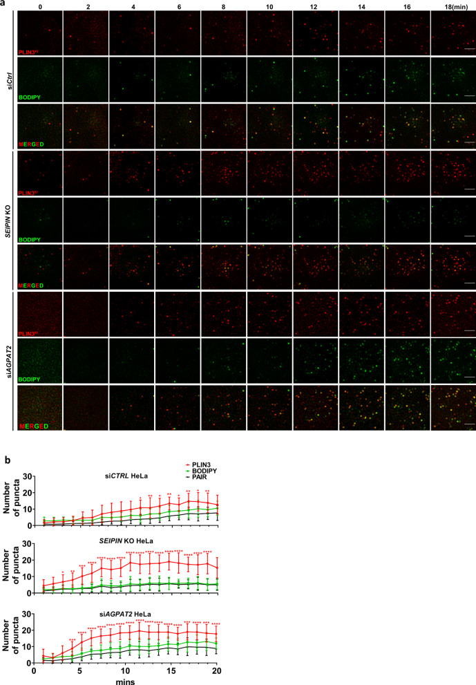
Fig. 2. AGPAT2 depletion impacted initial LD formation.
a Control, seipin knockout (KO) and AGPAT2 knockdown HeLa cells deficient were starved in 1%LPDS for 16 h. Representative images show the localization pattern of endogenous PLIN3 (mCherry-tagged) and BODIPY puncta every two minutes after oleate addition (400 µM). Bars: 5 μm. b The number of PLIN3 and BODIPY puncta in control, seipin KO and AGPAT2 knockdown HeLa cells at indicated time points. Pair indicates colocalization of PLIN3 and BODIPY puncta. (mean ± SD; ****p < 0.0001; ***p < 0.001; **p < 0.01; *p < 0.05, two-way ANOVA, n = 15–20 cells examined over three biologically independent experiments).

