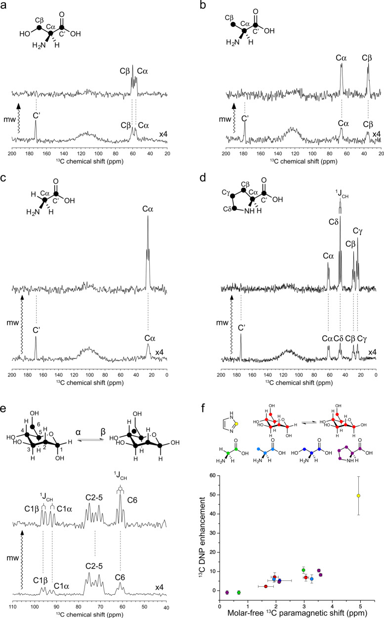
Fig. 2. Room-temperature ODNP NMR of small biological molecules in H2O at 9.4 T.
TEMPOL (100 mM) was used as the hyperpolarizing agent. The ODNP-enhanced 13C NMR spectra of U–13C serine (a), alanine (b), glycine (c), proline (d), and glucose (e) are scaled with the number of scans for visualizing directly the enhancement factor. All the microwave (mw)-off spectra in (a–e) are further scaled up by four-fold for the better visualization. The resolved 1JCH are showcased in spectral (d, e). f A positive trend of ODNP 13C enhancement over molar-free paramagnetic 13C NMR shift is visualized. The data on various molecules are presented in colors. The error of is defined as fitting error or standard deviation of values on carbons showing overlapping signals on ODNP spectra. The error of ODNP enhancement is determined from signal-to-noise ratio. More details about the error calculations can be found in “Methods”. Source data are provided as a Source Data file.

