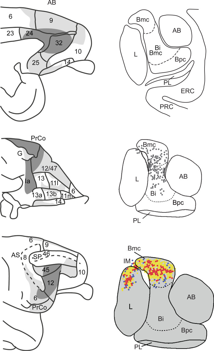
Fig. 1. Anatomical connections of the amygdala and prefrontal cortex in macaque monkeys.
Left. Schematic summary of macaque medial (top), orbital (middle) and lateral (bottom) frontal cortex regions in receipt of direct projections from the amygdala. Summary is based on anterograde tracers injected into the amygdala. The darker the shading, the greater the density of the terminal labeling. Numerals indicate cytoarchitectonic subdivisions. AS arcuate sulcus, SP principal sulcus, PrCo precentral opercular areas, G gustatory cortex, Ia agranular insular cortex. Adapted from [26]. Right. Coronal sections at the level of the mid-amygdala showing the typical layout of amygdala nuclei and the close proximity to neighboring entorhinal and perirhinal cortex (top), location of cells giving rise to projections to area 45 of VLPFC (middle), and locations of cortico-amygdala terminals received from area 45 (bottom). Adapted from Gerbella et al. [34, 234]. AB accessory basal nucleus, Bi basal nucleus intermediate division, Bmc basal nucleus magnocellular division, Bpc basal nucleus parvocellular division, ERC entorhinal cortex, IM intercalated masses, L lateral nucleus, PL paralaminar nucleus, PRC perirhinal cortex. Central and medial nuclei of the amygdala are not illustrated.

