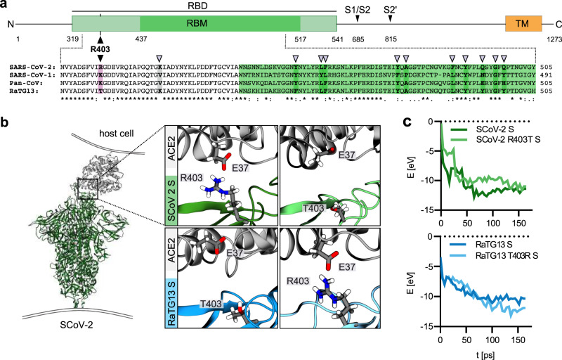
Fig. 1. Modelling of the interaction of Coronavirus Spike residue 403 with human ACE2.
a Schematic representation of the SARS-CoV-2 S protein (top panel), domains are indicated in different colours. Receptor binding domain (RBD), light green. Receptor binding motif (RBM), dark green. Transmembrane domain (TM), orange. R403, pink. S1/S2 and S2′ cleavage sites are indicated. Sequence alignment of SARS-CoV-2, SARS-CoV-1, Pan-CoV, and RaTG13 Spike RBD (bottom panel). Sequence conservation is indicated. grey arrows denote important residues for ACE2 binding. b Reactive force field simulation of SARS-CoV-2 Spike in complex with human ACE2 (PDB: 7KNB (https://www.rcsb.org/structure/7KNB)) (left panel) and focus on position 403 in SARS-CoV-2 S (R) or RaTG13 S (T) or respective exchange mutants at position 403 (right panel). c Exemplary energy curve of the reactive molecular dynamics simulation for SARS-CoV-2 S and SARS-CoV-2 S R403T (top panel) and RaTG13 and RaTG13 T430R spike with human ACE2 (bottom panel).

