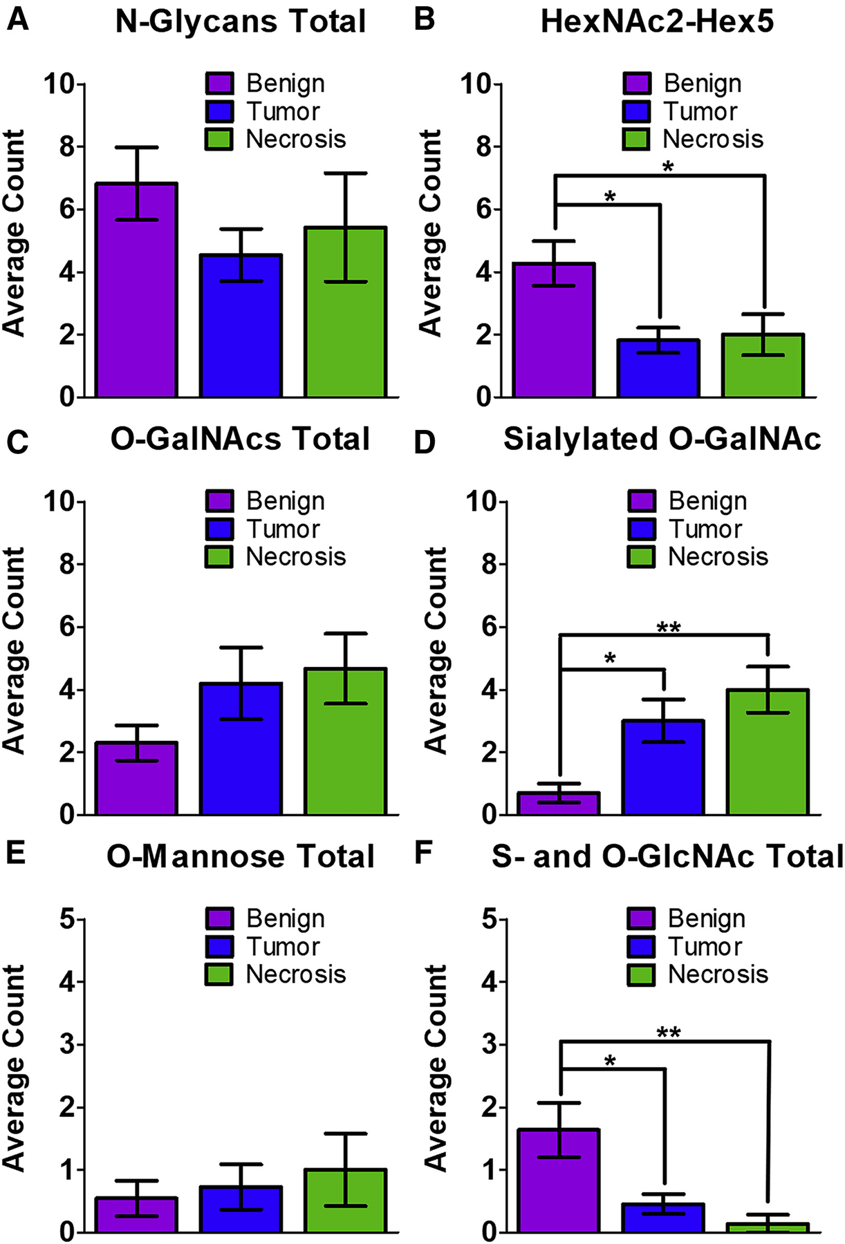
Figure 5. Statistical analysis of glycopeptide changes between samples.

For all samples, a three-way ANOVA test was performed in GraphPad PRISM, mean ± standard error of the mean, *p < 0.05, **p < 0.01.
(A) The total number of N-glycopeptides was not found to be significantly different between the three types of samples.
(B) The number of glycopeptides modified by HexNAc2-Hex5 was found to be significantly higher in the benign samples compared with both tumor and necrotic regions.
(C) The total number of O-GalNAcylated peptides was not was not found to be significantly changed between the three regions.
(D) The number of sialylated O-GalNAc glycopeptides was significantly increased in tumor and necrotic regions when compared with benign regions.
(E) The total number of O-mannosylated peptides was not was not found to be significantly changed between the three regions.
(F) S- and O-linked GlcNAcylated peptides were significantly decreased in both tumor and necrotic regions when compared with benign regions.
