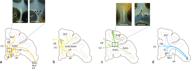
Fig. 2. Schematic demonstrating axons entering the white matter from ventral, medial, dorsal, and lateral PFC regions and branching to enter different pathways.
a Fibers exiting the OFC. Box indicates the inset of photomicrographs of the stalk and branching axons. b Fibers exiting the anterior cingulate cortex. c Fibers exiting the dorsolateral PFC. Box indicates the inset of photomicrographs of stalk and branching axons. d Fibers exiting the ventrolateral prefrontal cortex. AF amygdalofugal pathway, CB cingulum bundle, CC corpus callosum, EC external capsule, Extm extreme capsule, IC internal capsule, IL inferior longitudinal fasciculus, MLF medial longitudinal fasciculus, SLF superior longitudinal fasciculus, UF uncinate fasciculus.

