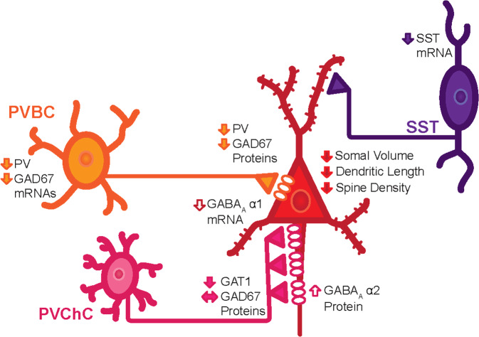
Fig. 2. Summary of cellular and molecular alterations in the DLPFC of individuals diagnosed with SZ identified through postmortem studies.
Pyramidal neurons in layer 3 of the DLPFC exhibit smaller somal volumes, shorter dendritic branches, and fewer dendritic spines. Two types of parvalbumin (PV) neurons provide inputs onto the perisomatic region of pyramidal neurons: PV basket cells (PVBC) and PV chandelier cells (PVChCs). The available data suggest that PVBC inputs exhibit lower levels of GAD67 protein, and putatively weaker GABA neurotransmission, in SZ. Postsynaptic to PVBC inputs are GABAA receptors enriched in the α1 subunit, and mRNA levels for this subunit are also lower in layer 3 pyramidal neurons in SZ, further contributing to weaker PVBC inhibitory inputs. In contrast, GAD67 levels appear to be unaltered in PVChC inputs, but these terminal cartridges exhibit lower protein levels of the GABA transporter, GAT1, and their synaptic targets, pyramidal neuron axon initial segments, exhibit higher protein levels of postsynaptic α2 subunit-containing GABAA receptors. In concert, these findings suggest that GABA neurotransmission is stronger at PVChC inputs in SZ. Finally, SST neurons, which appear to principally provide inputs onto the dendrites of pyramidal neurons in layers 2–3 of the DLPFC, exhibit lower levels of SST mRNA. Together, these alterations are thought to contribute to the neural substrate of cognitive dysfunction in SZ.

