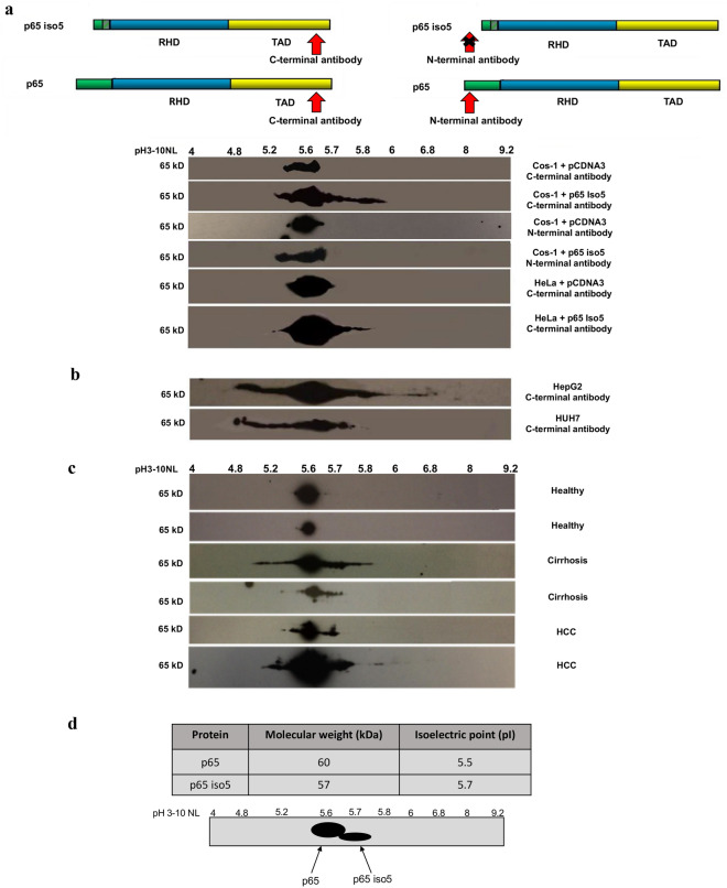
Figure 7.
p65 and p65 iso5 protein expression by 2-DE in human liver samples, transfected cell lines and human liver carcinoma cells. (a) Expression of p65 and p65 iso5 proteins by 2-DE in Cos-1 and HeLa cells transfected with p65 and p65 iso5 proteins with specific antibodies for C-terminal and N-terminal epitope region. (b) Expression of p65 and p65 iso5 proteins by 2-DE in HepG2 and HUH7 hepatocarcinoma cell lines. (c) Electrophoretic pattern of the p65 and p65 iso5 proteins on human liver of healthy, cirrhosis, and hepatocellular carcinoma (HCC) samples. The p65 antibody is specific for an epitope at C-terminal region that is present both in p65 and in p65 iso5 proteins. (d) Theoretical Molecular weight (Mw) and Isoelectric point (pI) of p65 and p65 iso5. The values were calculated using Compute pI/Mw tool (ExPASy) and do not take into consideration the possible post-translational modifications (PTMs). Schematic representation based on the hypothetical pI and Mw of the p65 and p65 iso5 protein pattern on 2D-PAGE. The full length images of 2-DE are available in the “Supplementary Information” file.

