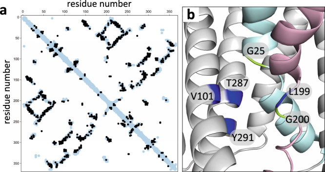
Fig. 1. GerAB resembles the l-alanine transporter GkApcT.

a Interaction matrix comparing evolutionary coupled (EC) residue pairs in GerAB (black circles) with residue pairs that are ≤5 Å apart in the GerAB model (light blue circles) derived from the GkApcT structure. 51% of EC pairs were within 5 Å of each other in the GerAB homology model. b Predicted l-alanine binding pocket in GerAB. TM segments 1 (cyan) and 6 (pink) are highlighted. G25 and G200 that are predicted to generate discontinuities in these helices are shown in green. Residues predicted to line the l-alanine binding pocket are indicated in dark blue. A side-by-side comparison to the l-alanine binding pocket in GkApcT is shown in Supplementary Fig. 1b.
