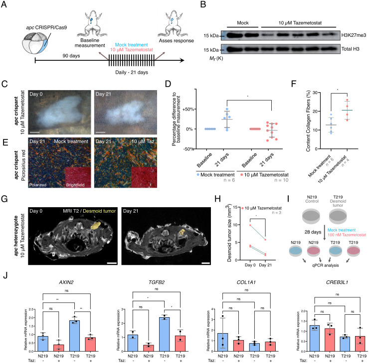
Fig. 5.
Ezh2 inhibition via Tazemetostat elicits a therapeutic response in desmoid tumors. (A and B) Tazemetostat-treated F0 apc crispants show a reduction in H3K27me3 levels in the liver (unaltered original scans of blots SI Appendix, Fig. S7). (C and D) Quantitatively, desmoid tumor size reveals stable or progressive disease in the control arm, while in Tazemetostat-exposed animals regression or stasis of tumors could be observed. Each data point represents one desmoid tumor; note that some animals developed multiple tumors (two-way ANOVA with repeated measurements; Dataset S6D; *P < 0.05). (E and F) Picrosirius Red staining with polarized light detection. The degree of alignment of collagen fibers is significantly increased in Tazemetostat-treated tumors when compared with mock-treated tumors (Student’s t test; Dataset S6D; *P < 0.05). (G and H) Desmoid tumors in apcMCR-Δ1/+ heterozygotes respond treatment with Tazemetostat (paired Student’s t test; Dataset S6D; *P < 0.05). (I) Treatment scheme of primary human desmoid tumor culture (T219) and paired normal cells derived from the fascia (N219) with Tazemetostat (100 nM) for 4 wk. (J) qRT-PCR analysis of AXIN2, TGFB2, COL1A1, and CREB3L1 expression in DMSO- (blue bars) or Tazemetostat (red bars)-treated cells (one-way ANOVA; Dataset S6D; *P < 0.05 **P < 0.01) (Gray scale bar, 1 mm; white scale bar, 3 mm).

