Figure 1.

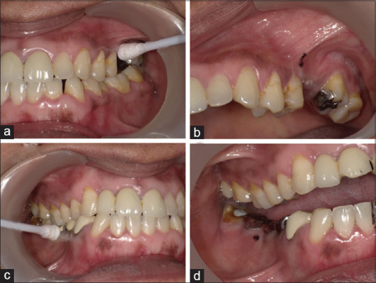
Localization and marking of the TrN lesions. (a and c) Pressure with cotton bud identifying a 3X3mm area on the mucobuccal fold and gingiva, (b and d) Site of the lesions marked with dye

Localization and marking of the TrN lesions. (a and c) Pressure with cotton bud identifying a 3X3mm area on the mucobuccal fold and gingiva, (b and d) Site of the lesions marked with dye