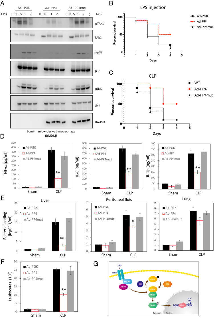
Fig. 4.
PP4 protected mice from CLP-induced polymicrobial sepsis. (A) BMDM cells infected with adenoviruses expressing EV (Ad-PGK) or gene encoding HA-PP4 WT (Ad-PP4) or HA-PP4 mutant (Ad-PP4mut) were stimulated with LPS for different time points and assessed by IB analysis with indicated antibodies. (B and C) Survival curves of Ad-PGK, Ad-PP4, or Ad-PP4mut infected mice following LPS injection (B) or CLP (C) (n = 10 per group per experiment). (D–F) Serum cytokines (D) and bacterial load (as CFU) in the lung, liver, and peritoneal fluid (E) and circulating leukocytes (CD11b+) (F) in Ad-PGK, Ad-PP4 or Ad-PP4mut infected mice were measured 24 h after sham or CLP (n = 10 per group per experiment). Data are the mean ± SE for each group. *P < 0.05, **P < 0.01 (by one-way ANOVA). (G) Model of TLR4/TRAF6/sNASP axis regulated by PP4. After bacterial infection (such as LPS), phosphorylation of sNASP dissociated from TRAF6 results in TRAF6 autoubiquitination and proinflammatory cytokine release. Then, PP4 is recruited to dephosphorylate sNASP to prevent persistent TRAF6 autoubiquitination and overwhelm proinflammatory cytokines production.

