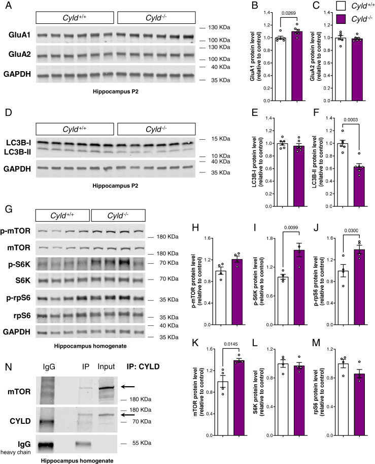
Fig. 5.
The loss of CYLD leads to an increase of the AMPA receptor subunit GluA1, dysregulates autophagic flux, and causes mTOR signaling up-regulation within the hippocampus. (A–C) Western blot analysis of the AMPA receptor subunits GluA1 and GluA2, each normalized to GAPDH in the hippocampal P2 fraction. (D–F) Western blot analysis of LC3B-I and LC3B-II, each normalized to GAPDH in the hippocampal P2 fraction. (A–G) Western blot analysis of mTOR signaling cascade and relative quantification in hippocampus homogenate. p-mTOR, p-S6K, and p-rpS6 were normalized to total mTOR, S6K, and rpS6, respectively. Total mTOR, total S6K, and total rpS6 were normalized to GAPDH. Lysates (20 µg proteins for A and D and 30 µg proteins for G) of Cyld−/− mice and Cyld+/+ controls were run on a 4 to 15% gradient gel. (H) CYLD immunoprecipitation from wild-type hippocampus homogenate run on a 4 to 15% gradient gel and blotted for mTOR and CYLD. For experiments in A through F, n = 6 for Cyld+/+ controls and n = 6 for Cyld−/− mice at P42. For experiments in G through M, n = 4 for Cyld+/+ controls and n = 4 for Cyld−/− mice at P42. Statistics were calculated by unpaired Student’s t test and show a significant increase in GluA1, a significant decrease in LC3B-II protein levels, and a significant increase in total mTOR, p-S6K, and p-rpS6 in Cyld−/− hippocampi. Graphs are mean ± SEM.

