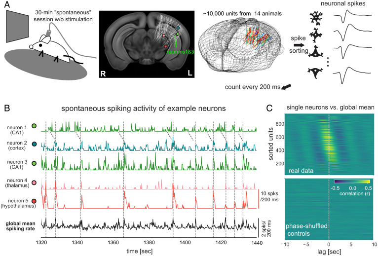
Fig. 1.
Neuronal population is engaged in global activity of the seconds timescale during immobile rest. (A) Illustration of the experimental setup of the Visual Coding – Neuropixels project that included a 30-min “spontaneous” session without any stimulation (Top Left). The locations of 6,171 channels on 79 probes from 14 mice were collapsed along the anterior-posterior direction and mapped onto a middle slice of a mouse brain template and also shown in a three-dimensional representation of the mouse brain (the second and third columns). (B) Spontaneous spiking rate of five example neurons from the hippocampus (CA1), visual cortex (VISal), thalamus (LP), and hypothalamus (ZI) of a representative mouse. Their locations were marked in (A). The neurons 1 and 3 were recorded by two channels with a distance of 14 μm. The Bottom trace is the global mean spiking rate of all 930 surveyed neurons. (C) Cross-correlation functions between individual neurons’ spiking rate and the global mean spiking rate (reference) during the stationary period from the representative mouse (Top). The same cross-correlation functions were computed after phase-shuffling the real data (Bottom).

