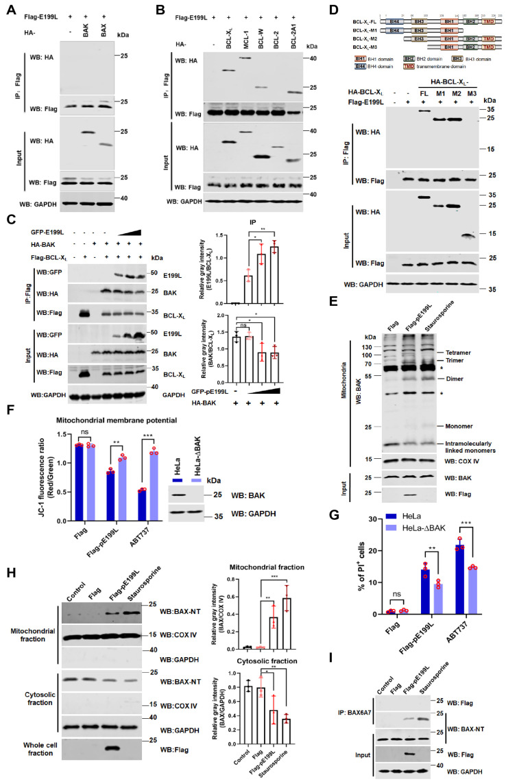
Figure 5.
ASFV pE199L promotes BAK activation by competing with BAK for BCL-XL. (A,B) Detection of the interaction of pE199L and apoptotic effectors (BAK and BAX) or anti-apoptotic proteins (BCL-XL, MCL-1, BCL-W, BCL-2, and BCL-2A1). HEK293T cells were transfected with pFlag-E199L alone or together with HA-BAK, BAX, BCL-XL, MCL-1, BCL-W, BCL-2, or BCL-2A1. Co-IP and Western blotting were performed to test their interactions. (C) Disruption of BCL-XL-BAK complex by pE199L. HEK293T cells were transfected with Flag-BCL-XL, HA-BAK, and pGFP-E199L as indicated, and the competitive interaction between proteins was detected by Co-IP and Western blotting (left). Ratios of pE199L or BAK and BCL-XL are shown as mean ± SD values from 3 independent replicates (right). The gray density of the protein bound was measured by Image J software. (D) Analysis of interaction between pE199L and the domain of BCL-XL. HEK293T cells were transfected with pFlag-E199L together with HA-BCL-XL and its mutants as indicated. The interactions were detected by Co-IP and Western blotting. (E) Induction of BAK homo-oligomerization by pE199L. Mitochondria from HeLa cells transfected with pFlag or pFlag-E199L were cross-linked with BMH as described in Materials and Methods. Asterisk (*) means a nonspecific band. (F) Detection of the mitochondrial membrane potential. HeLa or BAK-deficienct HeLa (HeLa-∆BAK) cells were transfected with pFlag-E199L or pFlag for 36 h or treated with ABT737 (10 μM) for 24 h as the positive control, stained with JC-1, and then quantified by flow cytometry as described in Materials and Methods. Western blotting was performed to validate BAK expression in both cell lines. (G) Analysis of BAK-dependent apoptosis induced by pE199L. The PI-labeled cells in HeLa and HeLa-∆BAK cells transfected with pFlag-E199L or pFlag were quantified by flow cytometry. The cells treated with ABT737 (10 μM) were used as a positive control. (H) Western blotting analysis of the translocation of BAX in the cytosol and mitochondrial fractions from HeLa cells transfected with pFlag-E199L or pFlag or treated with staurosporine as a positive control (left). The COX IV and GAPDH were respectively used as the internal control of mitochondrial or cytosolic fractions. Ratios of BAX and COX IV or GAPDH are shown as the mean ± SD values from 3 independent replicates (right). The gray density of the protein bound was measured by Image J software. (I) Western blotting analysis of the BAX activation in HeLa cells transfected with pFlag-E199L or pFlag. Cells were lysed and immunoprecipitated with the anti-BAX6A7 antibody. Equal amounts of the precipitated protein and cell lysates were immunoblotted with an anti-BAX-NT antibody. The cells treated with staurosporine were used as a positive control. The significance of differences between the groups was determined by the ordinary ANOVA test with Dunnett or 2-way ANOVA with Sidak (* p < 0.05, ** p < 0.01, *** p < 0.001).

