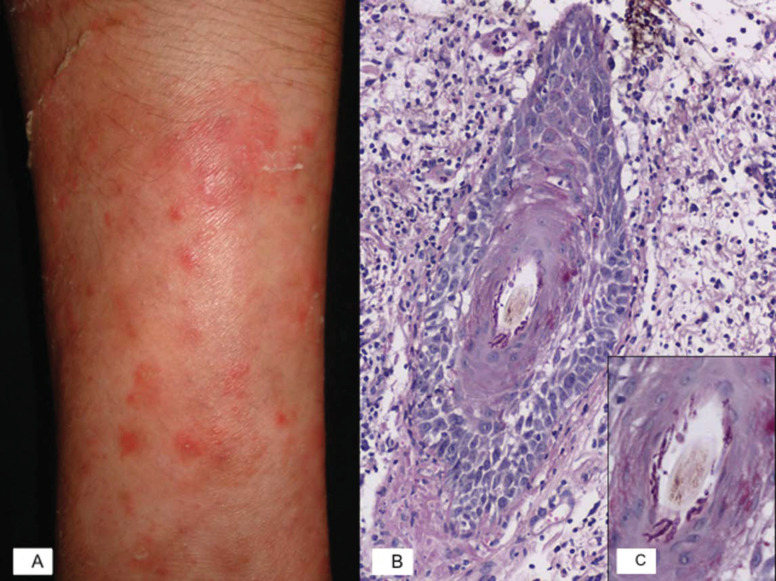
Figure 1.
Inflammatory tinea: Majocchi’s granuloma. (A) Majocchi granuloma characterized by erythematous nodules on the forearm. (B) Microphotography of a PAS-stained slide shows a vellus hair with adjacent mixed inflammatory infiltrate, epithelium spongiosis, exocytosis of neutrophils, and hyphae surrounding the hair shaft. (C) Closer view of the vellus hair showing septate hyphae within the follicular canal.

