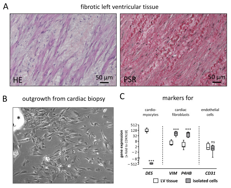
Figure 1.
Isolated cardiac cells derived from left ventricular tissue were characterized as fibroblasts. (A) Left ventricular (LV) tissue sections were stained using Hematoxylin & Eosin (HE) and Pico Sirius Red (PSR). In the HE staining, spindle-shaped cells—presumably cardiac fibroblasts—are visible in the fibrotic LV tissue. The cells are embedded within thick, curled fibers, which are confirmed as collagen fibers by PSR staining. (B) Spindle-shaped cells are outgrowing from a cardiac biopsy (upper left side, asterisk) and spreading over the cell culture plate. (C) To confirm the isolated cardiac cells as fibroblasts, the gene expression of LV tissue and isolated cardiac cells were compared regarding cell-type-specific markers. In the LV tissue, the muscle marker desmin (DES) is higher expressed, while vimentin (VIM) and prolyl 4-hydroxylase (P4HB) as markers for fibroblasts are lower expressed, compared to the isolated cardiac cells. A difference in the expression of the endothelial marker CD31 is not observed between LV tissue and isolated cardiac cells. For statistical comparisons, a t-test was used. *** p ≤ 0.001; ns, not significant.

