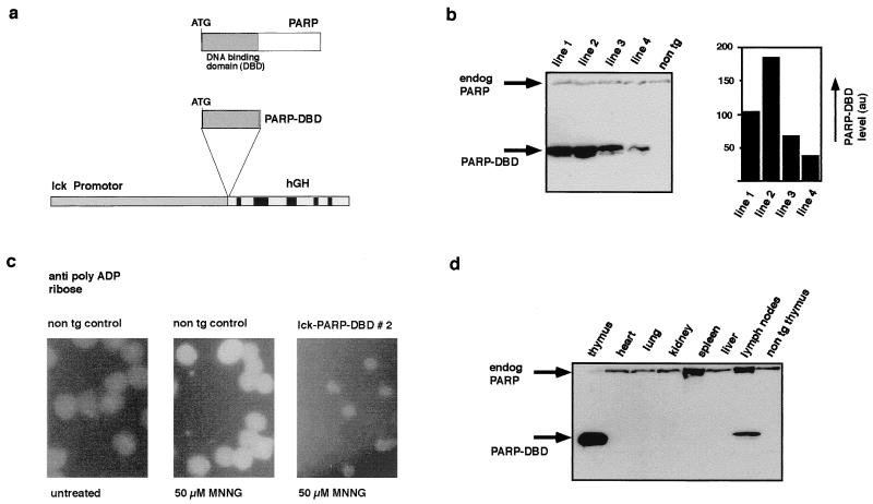
FIG. 1.
Introduction of a functionally active dominant negative mutant of PARP into the germ line of transgenic mice. (a) Schematic representation of the construct used to express the DBD of PARP in T cells of transgenic mice. (b) Expression of the lck-PARP DBD transgene in extracts from thymi of mice from four different transgenic lines (lck-PARP DBD lines 1, 2, 3, and 4). The level of transgene expression was measured by direct scanning of the ECL-treated membrane after blotting with a video camera and subsequent analysis by the AIDA software (Raytest, Straubenhardt, Germany) on a Fuji phosphorimager. PARP DBD expression was found to be higher in lines 1 and 2 than in lines 3 and 4. The PARP DBD protein was detected with the monoclonal antibody CII10, which recognizes the hPARP transgene and the endogenous mPARP. Relative expression levels are given in arbitrary units (au) obtained through the scanning procedure. (c) Dominant negative effect of the expression of the PARP DBD in T cells. Thymocytes from wild-type and lck-PARP DBD transgenic mice were treated with MNNG, and the formation of poly(ADP-ribose) was followed by immunofluorescence. Compared to nontransgenic (non tg) controls, thymocytes from lck-PARP DBD transgenic mice lack detectable polymer formation after MNNG treatment. (d) Expression level of the PARP DBD in different organs of a transgenic mouse of lck-PARP DBD line 2. As a control, an extract from a nontransgenic thymus was loaded. The PARP DBD protein was detected with the monoclonal antibody CII10, which recognizes the hPARP transgene and the endogenous mPARP. The relative amount of PARP DBD protein expression in splenocytes is detectable but very low (data not shown).

