
Figure 1.
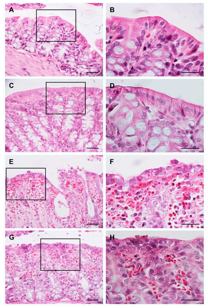
Evaluation of the morphology of the colon using the H&E stain. Images: (A,C,E,G) are at 40× magnification, images: (B,D,F,H) represent 100× magnification of the region marked with the rectangle in 40× images. Scale bar: 100 μm in all images.