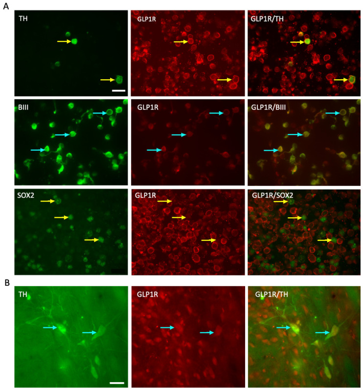
Figure 2.
GLP-1R expression on the E14 VM cells and dopaminergic neuronal graft in the striatum. (A) florescent images showing co-expression of GLP-1R (red) with TH+ cells (green), BIII tubulin+ cells (green) and SOX2+ cells (green) in E14 VM cells before grafting. (B) florescent images showing GLP-1R expression with TH+ cells in the grafted striatum after 13 weeks of transplantation (The arrow points to the co-localised points). Scale bar (A,B) = 20 μm. TH = tyrosine hydroxylase; GLP-1R = glucagon like peptide -1 receptor.

