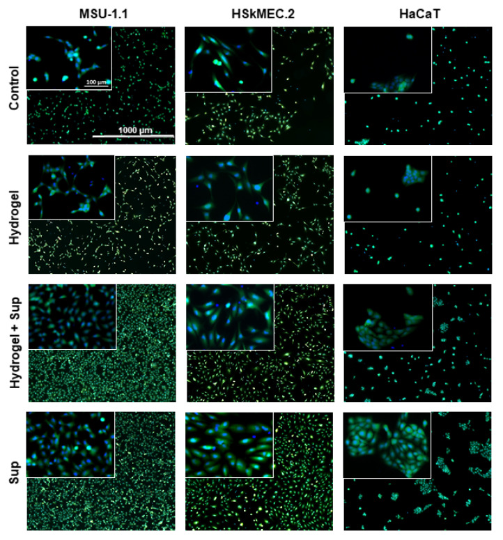
Figure 5.
Proliferative activity of skin-derived cells under hydrogel-released HATMSC2 trophic factors measured by Live/Dead assay. Fluorescent images (calcein-green and DAPI-blue) of MSU-1.1 (LH panel), HSkMEC.2 (middle panel) and HaCaT (RH panel) following three-day culture in serum-free medium and 1% O2 (untreated controls) and cells treated with empty hydrogel, supernatant-loaded hydrogel and 22 µg HATMSC2 supernatant protein. Inserts on the left top corners are 10× magnifications of the original images.

