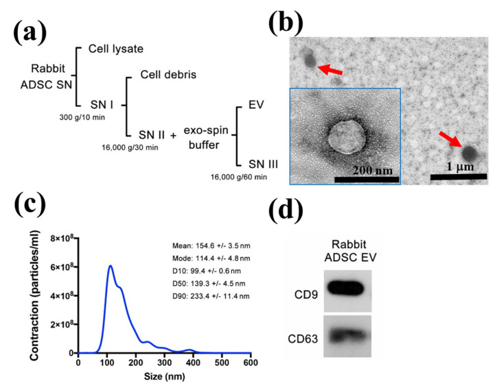
Figure 1.
Preparation and characterization of ADSC-EVs. (a) Flowchart demonstrating the isolation of EVs from the culture supernatant (SN) of ADSCs by differential centrifugation. (b) Rounded morphology and bilayer structure of ADSC-EVs was observed using TEM. (c) Size distribution was analyzed using NTA. (d) Western blotting to detect CD9 and CD63, specific surface markers of EVs.

